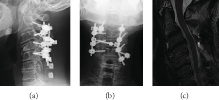
Figure 5.

(a, b) Postoperative X-ray. X-ray shows C2-5 posterior fusion. Anterior spondylolisthesis of the axis was corrected. (c) Postoperative MRI image. MRI image shows that decompression had been performed and that the central cord deficit had been improved.
