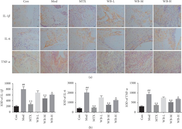
Figure 3.

(a) Representative immunohistochemistry images of IL-1α, IL-6, and TNF-α in each group. (b) IOD means of each group. Synovium tissue sections from ankle joints in each group were stained with anti-IL-1β, anti-IL-6, and anti-TNF-α. Original magnification 400x. ∗P < 0.05,∗∗P < 0.01 vs. model group. ##P < 0.01 vs. control group.
