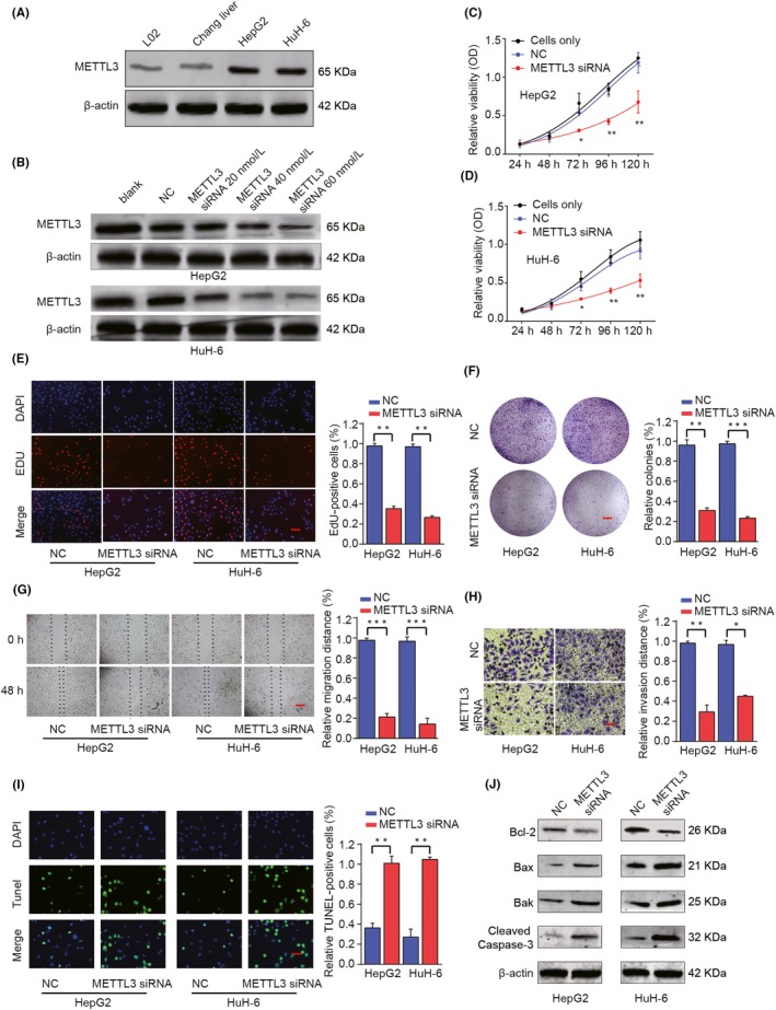
Figure 4.

Knockdown of METTL3 inhibits proliferation, migration and invasion of HB cells in vitro. A, The protein expression levels of METTL3 in HB cell lines (HepG2 and HuH‐6) and normal liver cell lines (L02 and Chang liver) were determined by Western blot analysis. HepG2 or HuH‐6 cells were transfected with negative control (NC) or different concentrations of METTL3 siRNA. B, The protein expression levels of METTL3 were analysed by Western blot 48 h later. C, D, CCK‐8 assay, (E) EDU staining (scale bars, 50 μm) and (F) colony formation assays (scale bars, 8 mm) were performed to determine the cell proliferation, DNA synthesis and colony formation in HepG2 or HuH‐6 cells. G, Wound‐healing assay (scale bars, 500 μm) and (H) Transwell assay (scale bars, 50 μm) were performed to assess the cell migration and invasion capacity. I, Representative micrographs and quantification of TUNEL‐positive signalling in the indicated assay (scale bars, 50 μm). J, Western blot analysis of Bcl‐2, Bax, Bak and cleaved caspase‐3 proteins in HepG2 and HuH‐6 cells. *P < .05, **P < .01, ***P < .001
