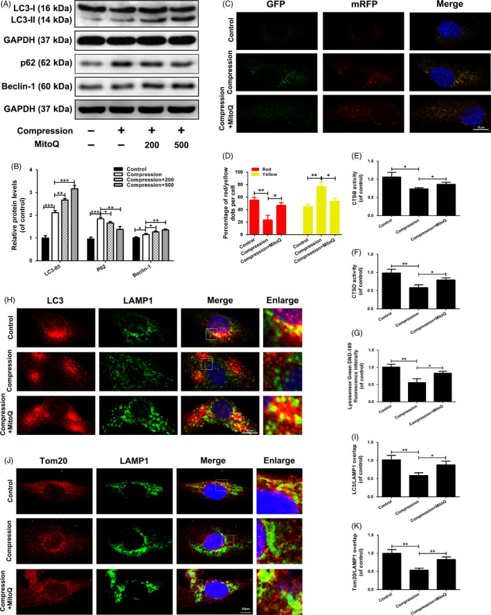
Figure 6.

MitoQ repairs defective mitophagic flux in human NP cells exposed to compression. (A‐B) The protein levels of LC3, p62 and Beclin‐1 in the human NP cells were measured by Western blotting. (C‐D) Representative images of the human NP cells expressing mRFP‐GFP‐LC3 were obtained by confocal microscopy. Scale bar: 10 μm. (E‐F) The activity of CTSB and CTSD in the human NP cells. (G) Lysosomal pH of human NP cells was determined by LysoSensor Green DND‐189 staining. (H‐I) The colocalization of LC3 and LAMP1 was examined by confocal microscopy. Scale bar: 10 μm. (J‐K) The colocalization of Tom20 and LAMP1 was examined by confocal microscopy. Scale bar: 10 μm. Data are represented as the mean ± SD. ***P < .001, **P < .01, *P < .05, n = 3
