
Figure 10.
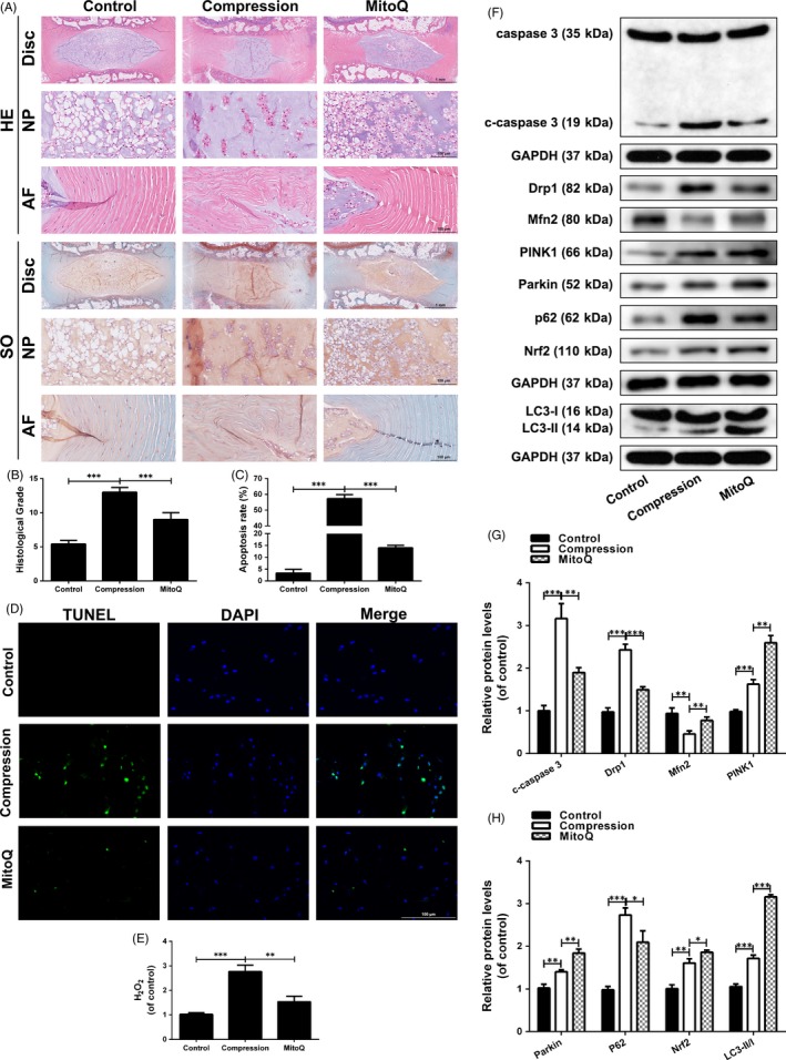
MitoQ ameliorates IDD development in an ex vivo model. The rat IVDs from compression group were cultured under compression treatment for 2 wk. The rat IVDs from MitoQ group were cultured under compression and MitoQ cotreatment for 2 wk. (A) HE and SO staining of the rat IVD tissues. (B) The histological scores of the rat IVD tissues according to histological grading scale. (C‐D) TUNEL staining and fluorescence microscope analysis were used to evaluate the apoptosis in the rat IVD tissues. Scale bar: 100 μm. (E) The content of H2O2 in the disc samples. (F‐H) The levels of caspase‐3, c‐caspase 3, Drp1, Mfn2, PINK1, Parkin, P62, Nrf2 and LC3 proteins in the rat IVD tissues were measured by Western blotting. Data are represented as the mean ± SD. ***P < .001, **P < .01, *P < .05, n = 6