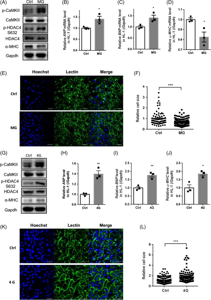
Figure 3.

Rotation‐simulated microgravity and hypergravity activated cardiomyocyte remodelling. A, Expression of CaMKII and its phosphorylation at Thr287 (p‐CaMKII), HDAC4 and its phosphorylation at Ser632 (p‐HDAC4) and α‐MHC in HL‐1 cells. B‐D, mRNA levels of ANP, BNP and α‐MHC in HL‐1 cells. E and F, Wheat germ agglutinin (WGA) staining was used to demarcate the boundaries of HL‐1 cells following rotation for 48 h. The cell area was analysed and quantified. Scale bar: 50 μm (n = 78 [Ctrl] and n = 151 [MG]). G, Expression of p‐CaMKII, p‐HDAC4 and α‐MHC following 4G hypergravity. H‐J, Analysis of ANP, BNP and α‐MHC mRNA levels following 4G hypergravity. K and L, WGA staining was used to demarcate the boundaries of HL‐1 cells following 4G centrifugation for 48 h. The cell area was analysed and quantified. Scale bar: 50 μm (n = 256 [Ctrl] and n = 125 [4G]). CaMKII, calcium/calmodulin‐dependent protein kinase II; HDAC4, histone deacetylase 4; α‐MHC, myosin heavy chain α. ANP, atrial natriuretic peptide; BNP, brain natriuretic peptide. Representative results of three independent experiments are shown. Data are shown as mean ± SEM; unpaired Student's t test, *P < .05, **P < .01 and ***P < .001
