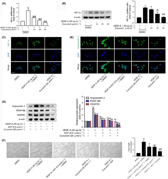
Figure 2.

HIF‐1α is involved in curcumol inhibition of liver sinusoidal endothelial cell angiogenesis. A, Real‐time PCR analysis of HIF‐1α. Data were expressed as percentage of control value (n = 4). B, Western blot analyses of HIF‐1α (n = 3). C and E, Immunofluorescence analysis of HIF‐1α and angiogenic cytokines (n = 3). Scale bar = 20 μm. D, Western blot analysis of angiogenic cytokines and ACF (0.5 μmol/L) was utilized to evaluate the function of HIF‐1α (n = 3). F, Tubulogenesis assay was visualized and quantified with ImageJ (n = 3). Scale bar = 500 μm. * P < .05, ** P < .01 and *** P < .005 versus control group; # P < .05, ## P < .01 and ### P < .005 versus model group
