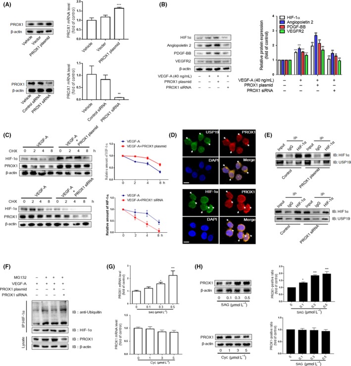
Figure 5.

PROX1 regulated by hedgehog signalling pathway maintains accumulation of HIF‐1α in liver sinusoidal endothelial cells (LSECs). A, Western blot and real‐time PCR analyses of efficiency of PROX1 siRNA and overexpression plasmid. B, Western blot analysis of HIF‐1α and angiogenic properties of LSECs treated with PROX1 siRNA and plasmid (n = 3). C, LSECs were infected with the vector plasmid and PROX1 siRNA or overexpression plasmid; then, the CHX (100 mg/mL) was used for the indicated time. Western blot analysis of HIF‐1α and PROX1 (n = 3). Means ± SD from three independent experiments were presented. D, The endogenous PROX1 and HIF‐1α/USP19 were visualized via confocal microscopy in primary LSECs (n = 3). Scale bar = 5 μm. E, Co‐IP assays were carried out with anti‐HIF‐1α and lgG as non‐specific control. The pull‐down lysate was detected using HIF‐1α and USP19 antibody. F, LSECs were treated with protease inhibitor MG132 (10 mg/mL) for 6 h after drug treatment. Co‐IP assays were conducted with anti‐HIF‐1α, followed by detection of the ubiquitin‐conjugated HIF‐1α with ubiquitin antibody. G and H, Western blot and real‐time PCR analyses of PROX1 in LSECs treated with Hh signalling agonist (SAG 0.3 μmol/L) or inhibitor (cyclopamine 3 μmol/L) (n = 3). * P < .05, ** P < .01 and *** P < .005 versus control group; # P < .05, ## P < .01 and ### P < .005 versus model group
