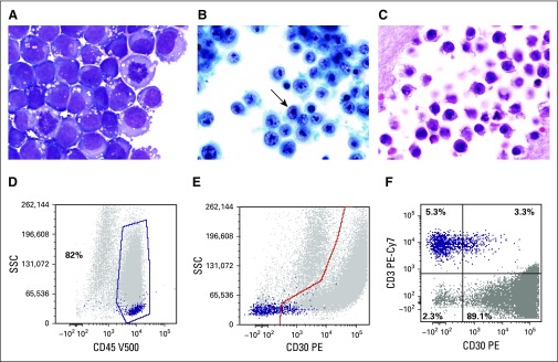
FIG 1.
Cytologic features of breast implant–associated anaplastic large-cell lymphoma (BIA-ALCL). (A) The neoplastic cells are large and pleomorphic with irregular nuclei, large prominent nucleoli, conspicuous mitotic activity, and moderate cytoplasm, which is basophilic and contains prominent vacuoles on air-dried preparations stained with a Romanowsky-type method (Giemsa, ×1,000). (B) The vesicular chromatin and nucleolar prominence and shape are better appreciated on the alcohol-fixed thin-layer preparation, which also contains some kidney-shaped hallmark cells (arrow; Papanicolaou, ×1,000). (C) Similar features are seen on the cell block preparation (hematoxylin and eosin, ×1,000). (D) Flow cytometric analysis of a BIA-ALCL effusion; in a histogram plotting CD45 versus side scatter, ALCL cells usually distribute in the lymphocyte (purple dots) and monocyte (gray dots) regions, that in this particular patient, represent 82% of cells in the specimen. (E) In a histogram plotting CD30 versus side scatter, CD30+ cells are distributed below the highlighted area (gray dots), whereas most lymphocytes are small and negative for CD30 (purple dots). (F) Histogram shows the distribution of selected (gated) cells in the histogram at top. It shows that 89.1% of gated cells express CD30, whereas 5.3% of cells are CD3+ lymphocytes but CD30−. It can be concluded that the aberrant cells (gray dots) are CD45+, CD30+, and CD3−, whereas the normal small T lymphocytes (purple dots) are CD45+, CD30−, and CD3+. PE-Cy7, phycoerythrin cyanine 7; SSC, side scatter; V500-A, Becton-Dickinson Horizon V500 (San Jose, CA) for flow cytometry.

