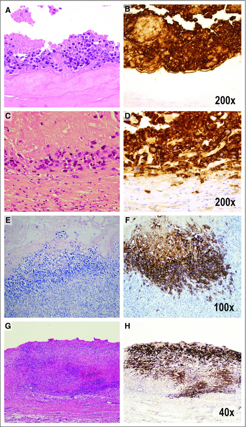
FIG 4.
Pathologic staging that reflects tumor progression from the luminal surface to the outer boundaries of the capsule. Pathologic stage T0 is not illustrated because it indicates the presence of tumor cells in the effusion, but not detected in the capsule. Pathologic stage T1 indicates (A) the lymphoma cells lie on the luminal side of the capsule, and CD30 highlights both viable and necrotic tumor cells, many of which are (B) ghosts in a background of granular material indicating cellular remnants. The tumor cells are insulated from the rest of the capsule by a dense acellular collagenous layer. Therefore, CD30 is much more extensive than the amount of identifiable viable tumor cells (A, hematoxylin and eosin [H&E]; B, CD30 immunohistochemistry, ×200). Pathologic stage T2 indicates few inflammatory cells admixed with deeper lymphoma cells (C, H&E; D, CD30 immunohistochemistry, ×200). Stage T3 indicates that numerous inflammatory cells surround lymphoma cells (E, H&E; F, CD30 immunohistochemistry, ×100). Stage 4 indicates that the tumor cells are beyond the boundaries of the fibrous capsule (G, H&E; H, CD30 immunohistochemistry, ×40).

