
Figure 3 .
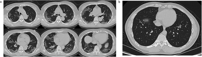
Typical CT images of 2019-nCoV pneumonia. (a) Low-dose high-resolution lung CT images from a 65-year-old man showing bilateral multiple lobar and segmental areas of ground-glass opacity, combined with interlobular septal thickening, on Day 8 after symptom onset. These are typical CT findings of severe cases. (b) A low-dose high-resolution lung CT image from a 34-year-old man showing patchy ground-glass opacity only in the anterior basal segment of right lower lobe on Day 3 after symptom onset. This is typical for mild cases.