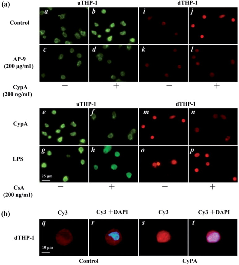
Fig. 6.

IF of NF-κB activity induced by CypA in the THP-1 cell. (a) The u and d THP-1 cells were precubated with control (a, b, i, j) and AP-9 (c, d, k, l), then stimulated with CypA (200 ng/mL, b, d, j, l), CypA/CSA (f, n), LPS (1 µg/mL, g, o) or LPS/CSA (h, p) respectively for 1h. The p50 protein of NF-κB activity was analysed by IF with FITC-conjugated anti-p50 antibody (green fluorescence) for the uTHP-1 and Cy3-conjugated anti-p50 antibody (red fluorescence) for the dTHP-1, respectively. CypA-induced nuclear translocation of NF-κB 2 h after treatment (b and h). Nuclear translocation of NF-κB induced by CypA was blockaded by AP-9 (d and l) or CSA (f and n) respectively. (b) Merged images of Cy3 and DAPI in the dTHP-1 were obtained using computer software. After CypA stimulating THP-1, the red color for NF-κB overlayed with the blue color for nucleus in cell nucleus under immunofluorescence microscopy, which showed plenty of p50 transducted into nucleus. The scale bars indicate 25 μm (a) and 10 μm (b). The data shown are representative of similar results from three independent experiments.
