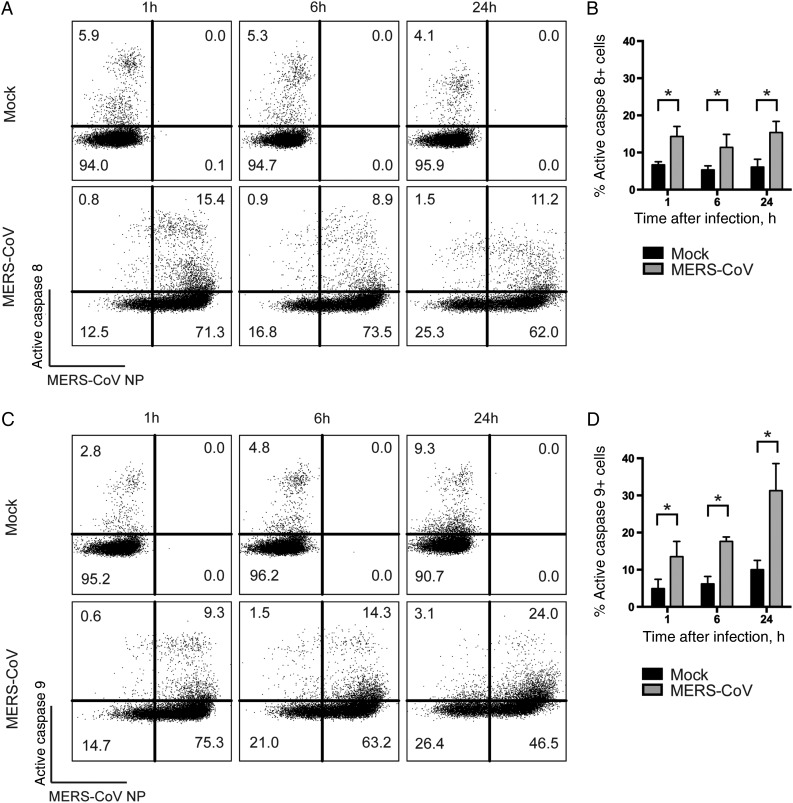
Figure 4.
The extrinsic and intrinsic caspase-dependent apoptosis pathways are activated in Middle East respiratory syndrome coronavirus (MERS-CoV)–infected T cells. T cells were infected with MERS-CoV at 2 50% tissue culture infective doses per cell. At the indicated time points, infected cells were labeled with MERS-CoV nucleoprotein (NP), as well as FITC-IETD-FMK (for caspase 8; A and B) and FITC-LEHD-FMK (for caspase 9; C and D) for 1 hour at 37°C. The average percentage of active caspase 8–positive (B) or active caspase 9–positive (D) cells among MERS-CoV NP–expressing cells from 3 different donors was illustrated. In all panels, bars and error bars represent means and standard deviations. Statistical analyses were performed using the Student t test. *P < .05.

