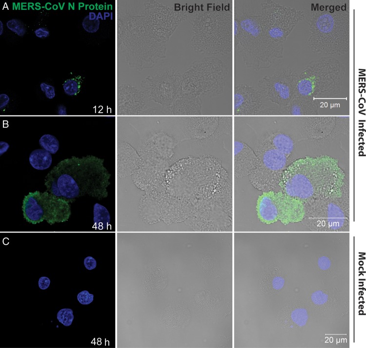
Figure 2.
Immunofluorescence staining of Middle East respiratory syndrome coronavirus (MERS-CoV) nucleocapsid protein (NP) in MERS-CoV–inoculated human monocyte–derived macrophages (MDMs). MDMs seeded on glass coverslips were inoculated with MERS-CoV at 2 tissue culture infective doses per cell or subjected to mock infection. Twelve hours (A) and 48 hours (B) after inoculation, cells were fixed, blocked, and permeabilized; incubated with anti-NP primary antibody for 1 hour; and stained with fluorescein isothiocyanate–conjugated secondary antibody for 1 hour. The slides were mounted with DAPI-containing mounting buffer and examined with a Carl Zeiss LSM 710 microscope. C, Findings for mock-inoculated MDMs that underwent the same procedure as that described for MERS-CoV–infected MDMs.

