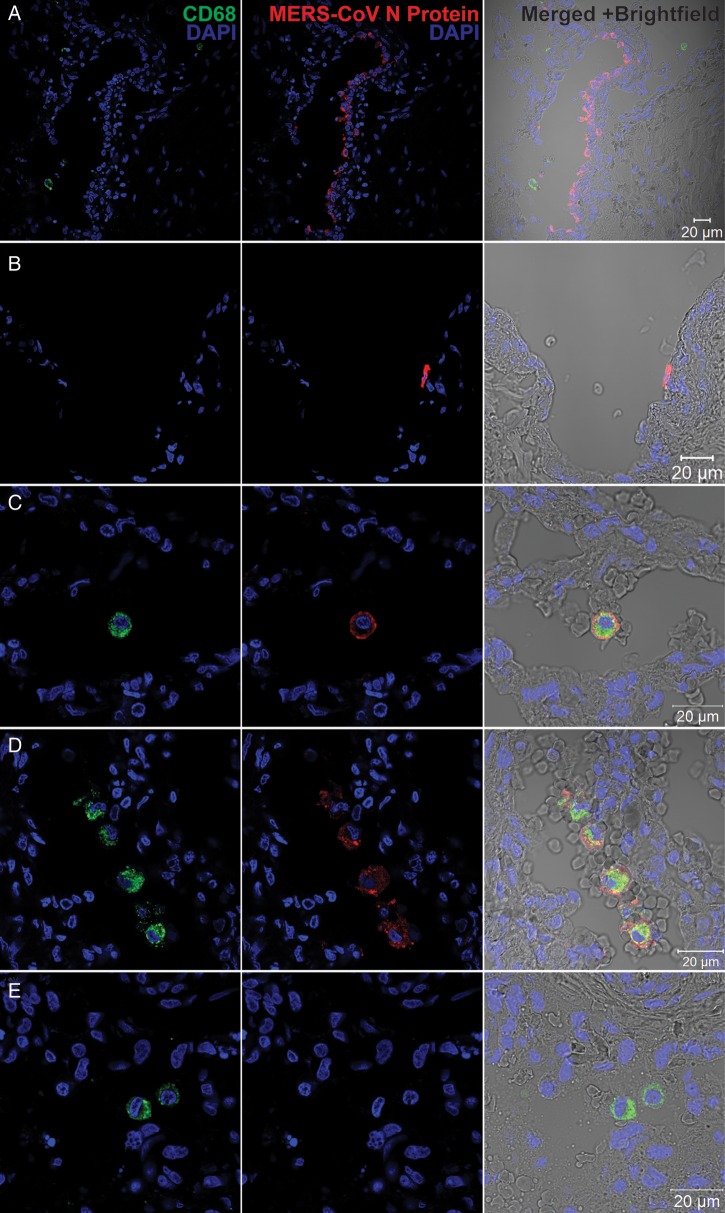
Figure 7.
Immunofluorescence double staining of Middle East respiratory syndrome coronavirus (MERS-CoV)–infected ex vivo lung tissue for MERS-CoV nucleocapsid protein (NP; red) and CD68 (green). Normal lung tissues were infected with MERS-CoV at 1 × 10⁷ tissue culture infective doses per milliliter or subjected to mock infection for 1 hour at 37°C. A total of 48 hours after infection, tissues were fixed, cryoprotected, and cryosectioned. Slides were sequentially stained with guinea pig anti-NP sera, Alexa 694 goat–anti-guinea pig immunoglobulin G (IgG), mouse anti-CD68 antibody and Alexa 488 goat–anti-mouse IgG. Cell nuclei were counterstained by DAPI in the mounting buffer. Images were captured with a Carl Zeiss LSM 710 microscope. Immunoreactivity to NP was detected in epithelial cells of terminal bronchiole (A) and vascular endothelial (B) cells. C and D, Colocalization of MERS-CoV NP and CD68. E, A representative image of mock-infected lung tissue.

