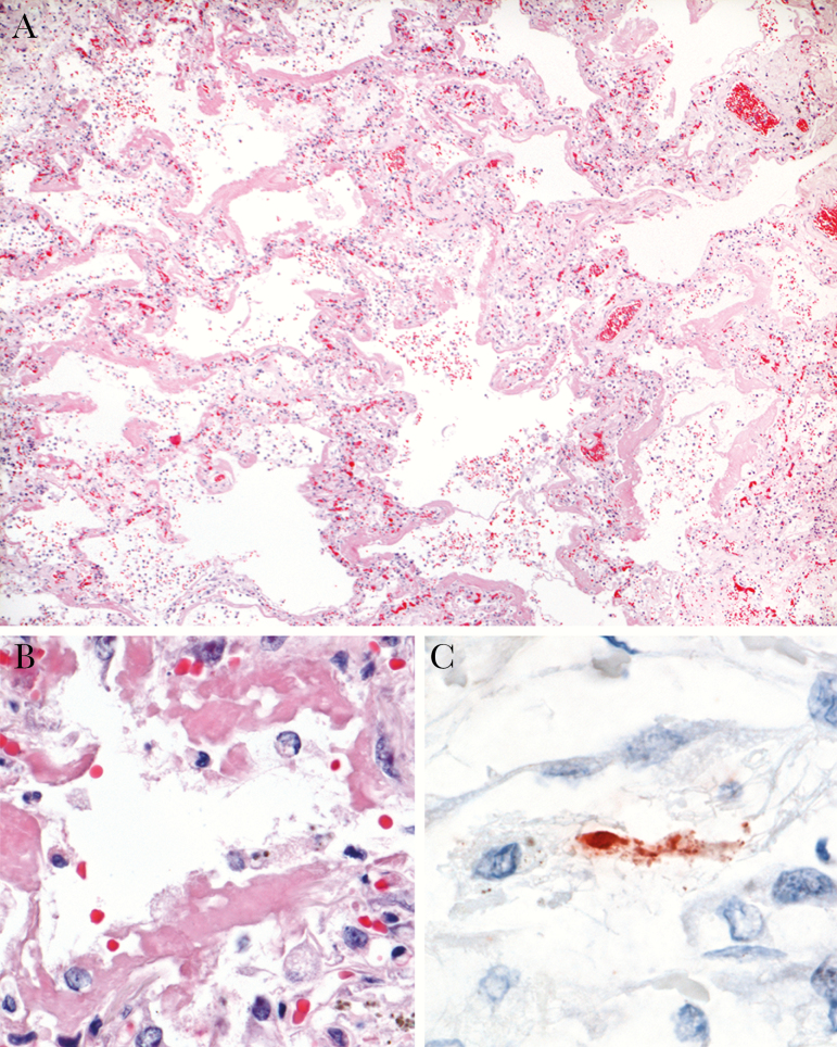
Figure 2.
Pulmonary immunohistochemistry and histopathology of the patient with hPPMV-1/NL/579/2003 infection. A, Diffuse alveolar damage, characterized by thickening of alveolar septa, flooding of alveolar lumina, and presence of hyaline membranes (hematoxylin and eosin [H&E] staining; original magnification ×4.) B, Higher magnification of A, showing single pulmonary alveole, where epithelial lining is replaced by hyaline membranes (H&E; original magnification ×40.) C, Avian paramyxovirus type 1 antigen visible as granular staining in cytoplasm of degenerate cell, suggestive of alveolar epithelial cell (immunoperoxidase stain for pigeon paramyxovirus type 1 [PPMV-1]; original magnification ×100.).

