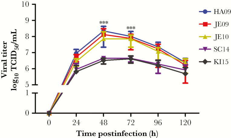
Figure 4.
Replication kinetics of A(H1N1)pdm09 viruses in porcine bronchial epithelial cells (PBECs). PBEC cultures were apically infected with pdmH1N1 isolates at infection dose of 104 TCID50/filter, at 37°C. After 1 hour, samples were washed 3 times with phosphate-buffered saline to remove free virions. Supernatants were collected at 24, 48, 72, 96, and 120 hours postinfection and analyzed for infectivity with TCID50 assay. At 48 and 72 hours postinfection, the differences were >10-fold and highly significant between viruses from 2009–2010 and viruses from 2014–2015. The results were shown as mean ± standard deviation of samples of 3 independent experiments, each with 3 replicates. Significance (***P < .001) was analyzed with Tukey multiple comparison test by using GraphPad Prism 5 software. Abbreviation: TCID50, tissue culture infectious dose 50.

