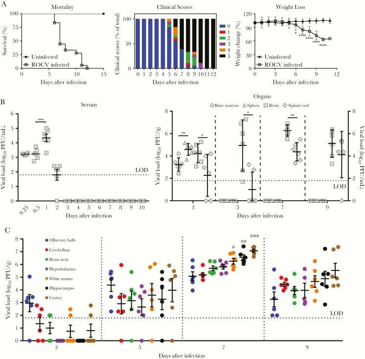
Figure 1.
Viral burden of Rocio virus (ROCV) replication in C57BL/6 mice. Six-week-old female C57BL/6 mice were inoculated intraperitoneally with 2.8 × 10⁶ plaque-forming units (PFU) of Rocio virus (ROCV; 20 mice/group). A, Survival rates (left), the percentage of the total clinical score (middle), and the percentage change in body weight (right) were determined up to 21 days after infection. B, Viral load in serum (left) and organ (right) specimens. C, Viral load in the major brain regions. Virus titers were determined by the plaque assay on BHK-21 cells and are presented as PFU per gram of tissue (for all tissue specimens) or PFU per milliliter (for serum or bone marrow specimens). #Statistically significant difference as compared to the olfactory bulb (P < .01) and cerebellum (P < .01). ##Statistically significant difference as compared to the olfactory bulb (P < .0001), cerebellum (P < .001), and brain stem (P < .05). ###Statistically significant difference as compared to the olfactory bulb (P < .0001), cerebellum (P < .0001), brain stem (P < .001), and hypothalamus (P < .01). None of the differences among the cortex, hippocampus, and white matter were statistically significant. In panels B and C, the horizontal dotted lines correspond to the limit of detection (LOD) of the plaque assay. *P < .05, **P < .01, ***P < .001, and ****P < .0001, by the Student t test (A), the Mann-Whitney test (B), or 1-way analysis of variance with the Dunnett multiple comparisons test (C).

