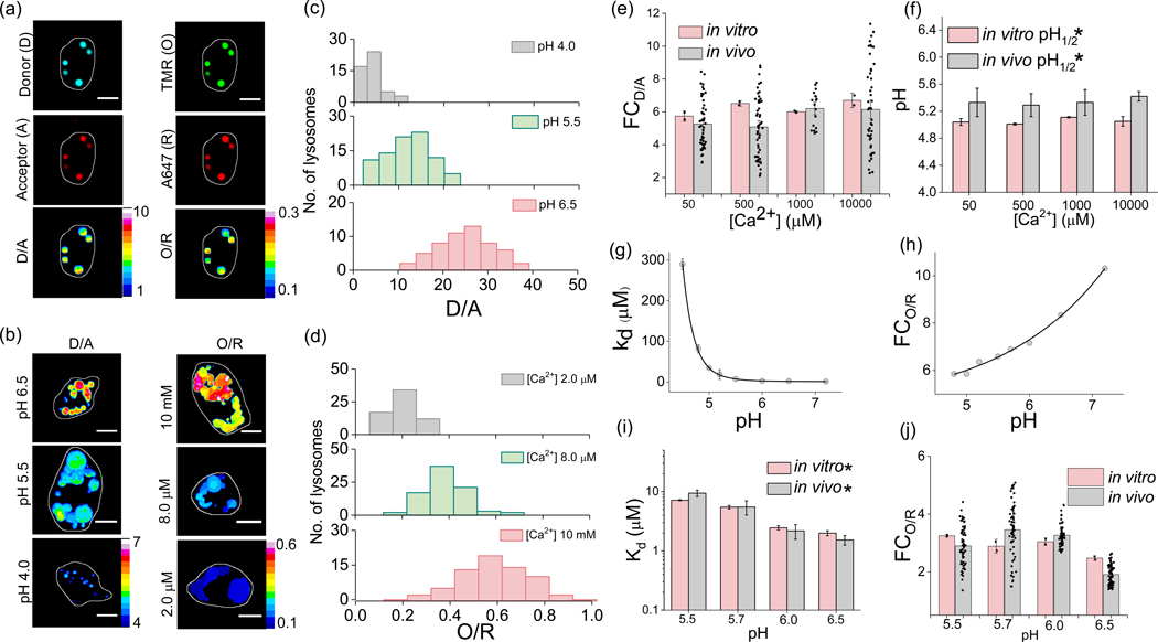
Figure-2: In vivo sensing characteristics of CalipHluorLy.
(a) Representative CalipHluorLy labelled coelomocytes imaged in the donor (D,i), acceptor (A,ii), Rhod-5F (O, iii) and Alexa647 (R, iv) channels. D/A (v) and O/R (vi) are the corresponding pixel-wise pseudocolor images. (b) Representative pseudo colored D/A and O/R maps of coelomocytes clamped at indicated pH and free [Ca2+] (c) Distribution of D/A ratios of ≥ 50 endosomes clamped at the indicated pH (n = 10 cells). (d) Distribution of O/R ratios of ≥ 50 endosomes clamped at different indicated free [Ca2+] (n = 10 cells). Comparison of (e) fold change of D/A ratios from pH 4 to 6.5 and; (f) pH1/2 from pH 4 to 6.5 of CalipHluorLy at different [Ca2+] obtained in vitro (peach) and in vivo (gray). CalipHluorLy (g) Dissociation constant Kd (μM) and (h) fold change of O/R as a function of pH. Comparison of (i) fold change in O/R ratio from 1 μM to 10 mM [Ca2+] and (j) Dissociation constant Kd (μM) of CalipHluorLy at the indicated pH obtained in vitro (peach) and in vivo (gray). Scale bars, 5 μm. Data represent mean ± s.e.m. * Error is obtained from the non-exponential fit. Experiments were repeated thrice independently with similar results.

