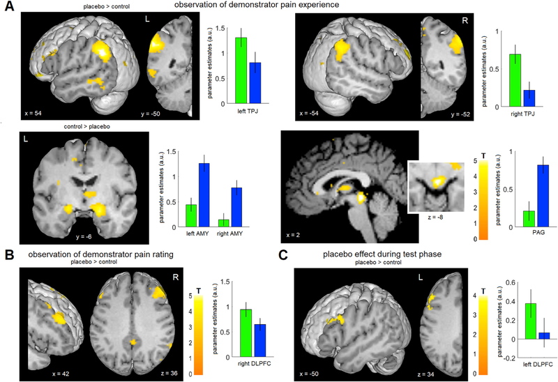
Fig. 3. Brain regions associated with observational learning and placebo hypoalgesia.
(A) While observing the pain experience of the demonstrator, participants showed a higher activation in the bilateral TPJ during the placebo as compared to the control condition. In the reverse contrast, a higher activation of the bilateral amygdalae and the PAG was observed. See also Fig. S1. (B) While observing the pain rating of the demonstrator, participants showed a higher activation in the right DLPFC in the placebo condition. (C) During the test phase, a higher activation of the left DLPFC was observed in the placebo condition. For visualization purposes, a voxels threshold of P < 0.005 uncorrected was used for the figure. Bar graphs indicate parameter estimates ± SEM within the ROIs for placebo (green) and control (blue).

