
Fig. 1.
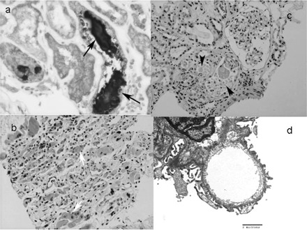
(a) The kidney from case 1. The renal tubule showed prominent cytoplasmic wasting pigment deposition with lymphocytes. The casts in the tubules (arrow) are positive for myoglobin (immunohistochemical stain, 200×). (b) The skeletal muscles from case 2. Myoid giant cells (white arrow) and interstitial ecstatic lymphovascular structures with inflammatory cell infiltration are evident (HE stain, 100×). (c) The kidney from case 2. There are several Mallory body-like casts (arrowhead) in dilated lumens with some sloughed and flattened renal tubular epithelial cells, indicating rhabdomyolysis and acute tubular necrosis. Interstitial infiltration of mononuclear cells is evident. There was a mild increase in mesangial cellularity and matrix (HE, 200×). (d) The glomerulus of case 2 (electron microscopy). Intact basement membrane surrounded by podocytes (×11 500). There was no immune complex deposition or virus particles.