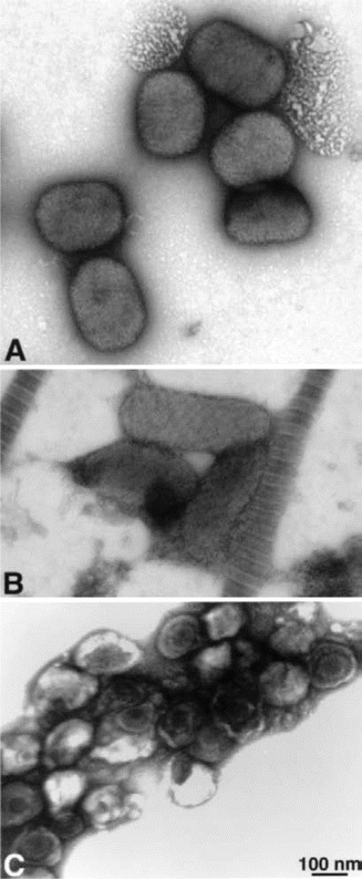
Figure 3.

Electron microscopy of negatively stained viruses. A, Viruses in the Orthopox genus (e.g., variola, vaccinia, monkeypox) and viruses in the Molluscipox genus (e.g., molluscum contagiosum) are morphologically similar. They are large, brick-shaped particles, ∼200 nm × 250–300 nm in size. Depending on the number and configuration of membranes around the particles, the surfaces may appear somewhat rough or with deep ridges. B, Parapox viruses (e.g., orf and Milker nodule virus) are slimmer and more elongated, 140–170 nm × 220–300 nm in size. The surface of these viruses frequently appears to have criss-crossed striations, like rope wrapped diagonally around the particle. C, Herpesviruses (e.g., varicella-zoster virus, human herpes virus, or herpes simplex virus) are 120–200 nm in diameter, with icosahedral nucleocapsids of ∼100 nm in diameter. Smallpox or variola can be rapidly (in 10–20 min) and easily distinguished morphologically from herpesviruses and parapoxviruses by negatively staining vesicle fluid. Electron photomicrograph is courtesy of Dr. Sara E. Miller.
