
Fig. 1.
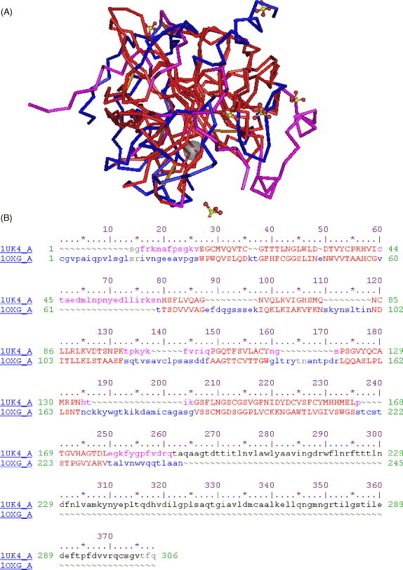
Structural similarity between the SARS coronavirus (SARS-CoV) main protease (3CLpro) and bovine α-chymotrypsin. (A) Three-dimensional structural superimposition between the HIV-1 protease (accession number in the Protein Data Bank: 1UK4) and α-chymotrypsin (accession number: 1OXG). The residues significantly aligned and corresponding to the catalytic site of both molecules are shown in red. The other regions of the catalytic domains of 3CLpro are shown in violet. Unaligned regions of α-chymotrypsin are shown in blue. (B) Sequence alignment corresponding to the structural alignment in (A). The colours of the residues in (B) strictly correspond to those of (A). This alignment was obtained using the VAST algorithm and visualised using the Cn3d 4.1 program (www.ncbi.nlm.nih.gov).