
Figure 1.
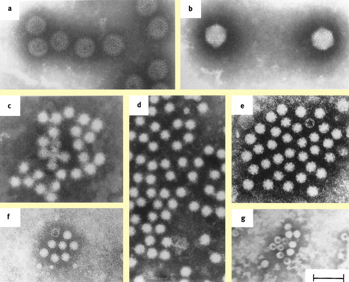
Electron micrographs showing. a rotavirus. b enteric adenovirus. c Norwalk-like virus. d calicivirus. e astrovirus. f enterovirus. g parvovirus. (Negative staining with 3% phosphotungstate, pH 6.3; bar 100 nm). (By courtesy Dr J Gray (a–d, f, g) and Dr J Kurtz (e)).Source: Zuckerman A, Banatvala J, Pattison J, eds. Principles and practice of clinical virology. 4th edn. Chichester: Wiley, 2000 © John Wiley & Sons Limited. Reproduced with permission.)