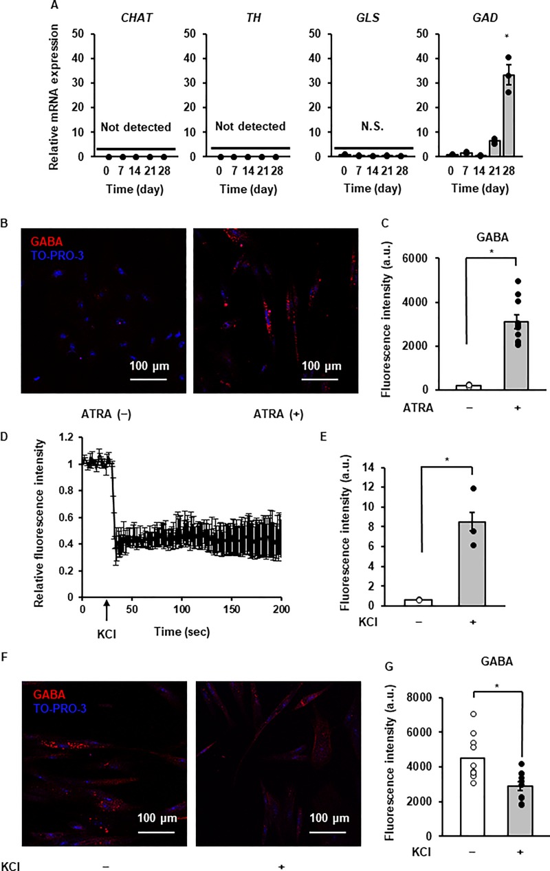
Fig 4. Neurotransmitter synthesis and secretion of ATRA-treated canine DFATs.
(A) Time-dependent changes in the mRNA expression of enzymes for neurotransmitter synthesis after ATRA treatment. Data are shown as mean ± standard error of three independent experiments. *P < 0.05, compared with 0 day. (B) Representative images of GABA (red) and TO-PRO-3 (blue; nuclei) with (right panel) or without (left panel; control) ATRA treatment after 28 days. (C) Fluorescence intensity of GABA (n = 10 cells, randomly selected ×20 fields from triplicate samples) with or without ATRA treatment after 28 days. Data are shown as mean ± standard error of three independent experiments. *P < 0.05. (D) Destaining of fluorescent false neurotransmitter stimulated by KCl (50 mM) in ATRA-treated DFATs. (E) Fluorescence intensity of fluorescent false neurotransmitter in the culture supernatant of cells treated with or without KCl (50 mM). Data are shown as the mean ± standard error of three independent experiments. *P < 0.05. (F) Representative images of GABA destaining in ATRA-treated DFATs treated with (right) or without (left) KCl (50 mM). (F) Fluorescence intensity of GABA (n = 10 cells, randomly selected ×20 fields from triplicate samples) with or without KCl treatment after 28 days. Data are shown as the mean ± standard error of three independent experiments. *P < 0.05.

