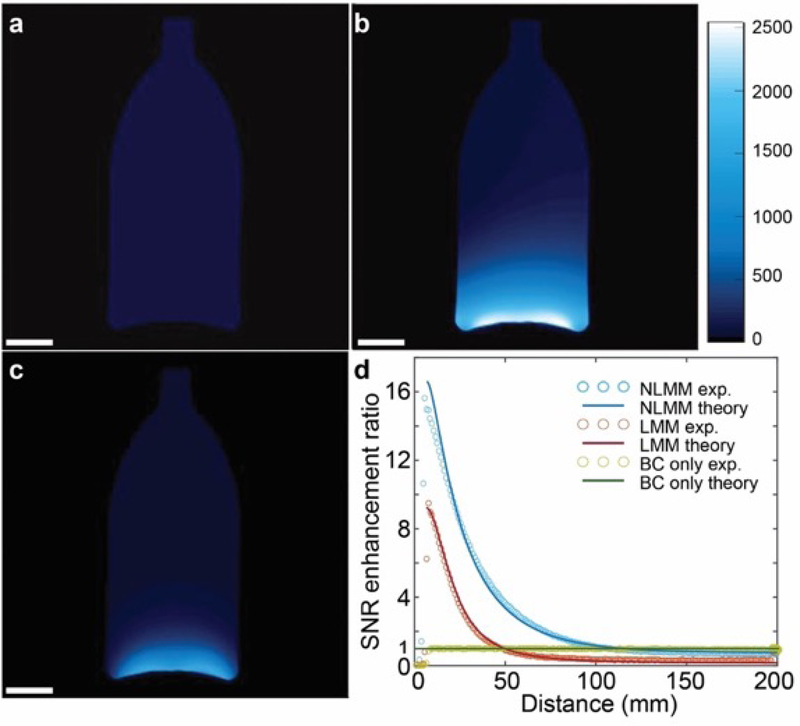
Figure 3.
3T MRI imaging of mineral oil phantom. Gradient echo imaging employed. a) Image captured by the body coil in absence of metamaterials. b) Image captured by the body coil in presence of NLMMs. c) Image captured by the body coil in presence of LMMs. d) Comparison of the SNR enhancement ratio for the nonlinear and linear MMs at the center of the phantom. Scale bars in a), b) and c) are 3 cm.

