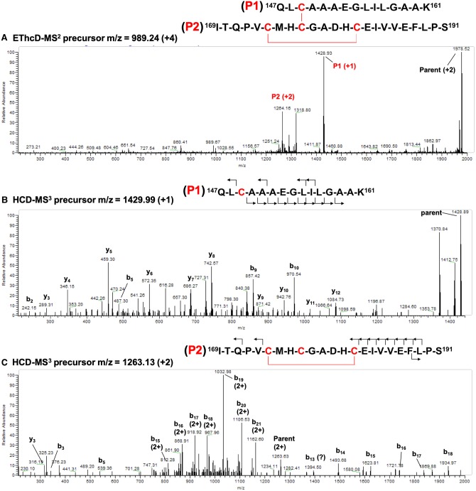
Figure 5. Mass spectra of the disulfide cross-linked peptide of oxidized H-NOX.
(A) EThcD-MS2 spectrum of m/z 989.24 (+4). (B) HCD-MS3 spectra of the P1 peptide with m/z 1429.99 (+1) and (C) the P2 peptide with m/z 1263.13 (+2). The peptide sequences with the observed fragment ions are shown above each spectrum with Cys residues and disulfide bonds indicated in red.

