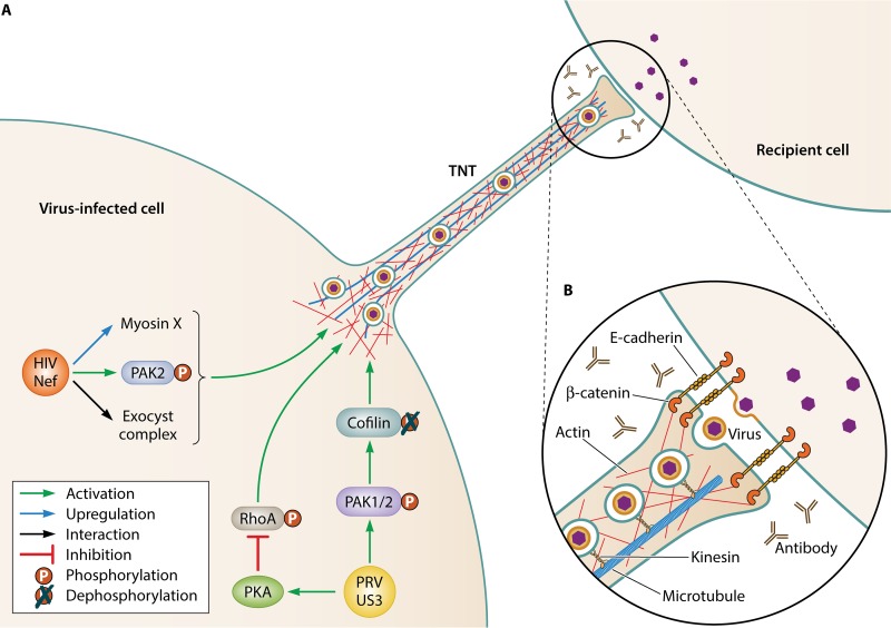
FIG 1.
(A) Schematic representation of two of the best characterized models of virus-induced TNT formation: Nef-induced TNT formation in HIV-infected macrophages and US3-induced TNT formation in PRV-infected epithelial cells. Both types of TNTs have been shown to carry virus particles in vesicles (see, for example, references 17 and 43). Some of the molecular players that have been shown to be involved in these two types of TNT formation are indicated. More detailed information on their role is provided in the present study. (B) An inset image shows a schematic representation of what is known about the contact area between a PRV US3-induced TNT and a recipient cell based on electron microscopy and confocal microscopy analyses (based on reference 17).

