. 2020 Mar 31;9:e49392. doi: 10.7554/eLife.49392

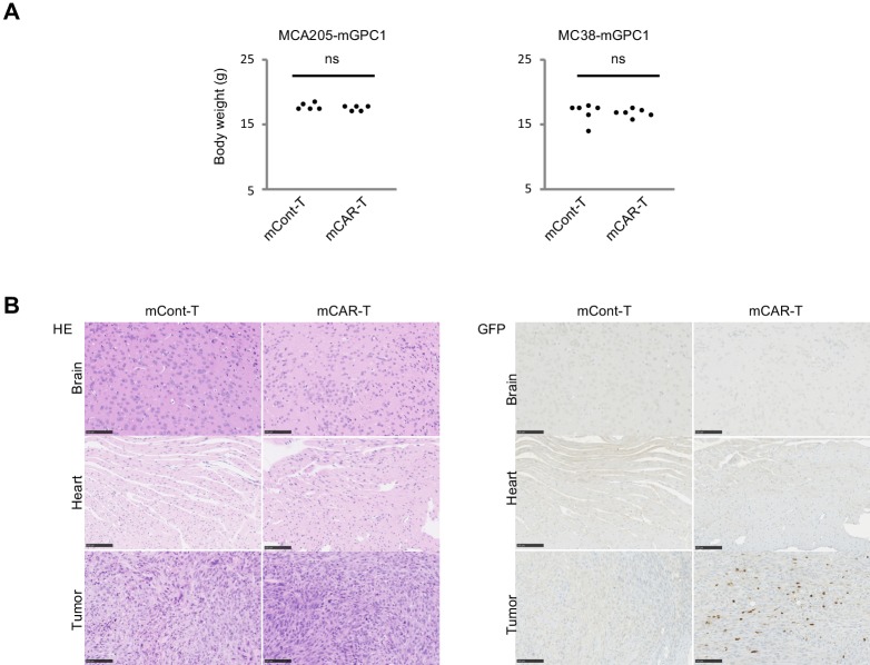
Figure 5. GPC1-specific mCAR-T cells showed no obvious adverse effects on normal tissues in vivo.

(A) Body weight of mCAR-T cells or mCont-T cells injected mice bearing MC38-mGPC1 (right panel) or MCA205-GPC1 (left panel) was measured 12 days after mCAR-T cell or mCont-T cell administration. (B) Representative staining of HE (left panel) and IHC for injected GFP+ T cells detected by anti-GFP Ab (right panel) in mouse normal tissues are shown. The data of other normal tissues are shown in Figure 5-figure supplements 1 and 2.

Figure 5.

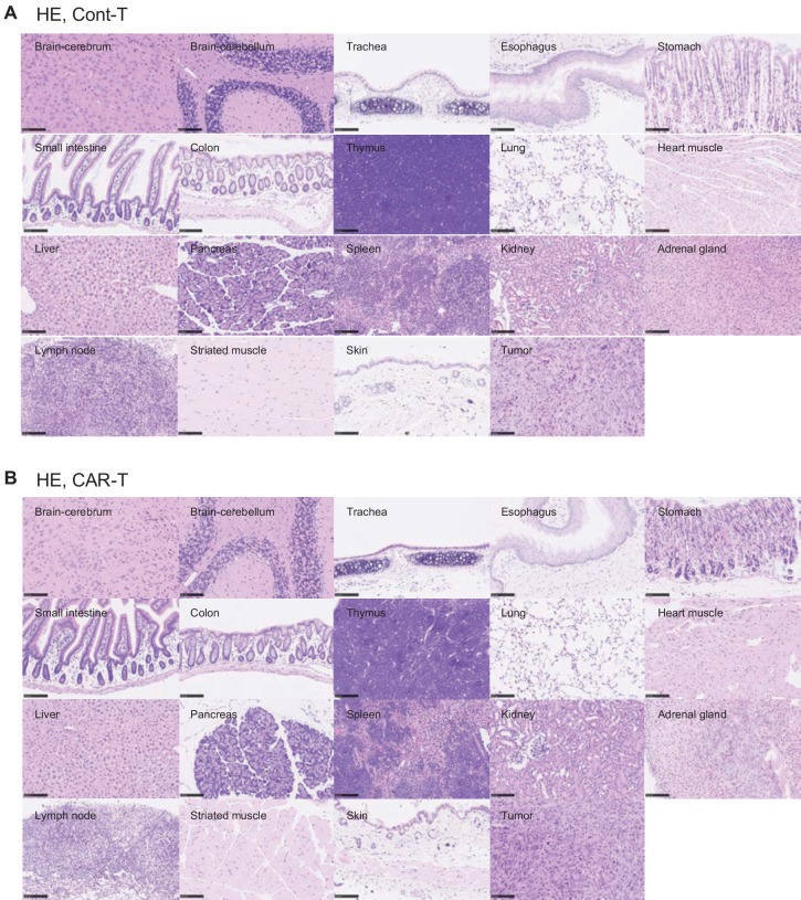
Figure 5—figure supplement 1. Tissues damages were not detected in mouse normal tissues.

Figure 5—figure supplement 1.

HE staining was performed on normal tissues from MC38-mGPC1-bearing mouse treated with mCont-T cells (A) or mCAR-T cells (B). Scale bar, 100 μm.
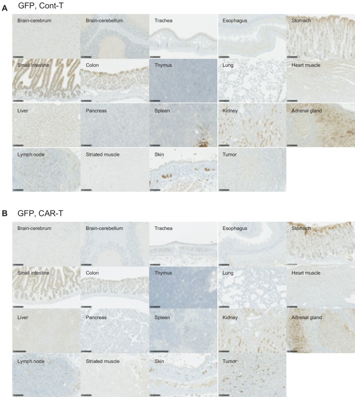
Figure 5—figure supplement 2. Only few GFP-positive mCAR-T cells infiltrated mouse normal tissues.

Figure 5—figure supplement 2.

IHC staining of GFP was performed on normal tissues from the MC38-mGPC1-bearing mouse treated with mCont-T cells (A) or mCAR-T cells (B). Scale bar, 100 μm.