. 2020 Mar 31;9:e48964. doi: 10.7554/eLife.48964

Figure 1. Reduction of Fibronectin matrix enhances neural tube convergence but abrogates bilaterally symmetric paraxial mesoderm morphogenesis.

(A) A schematic of the zebrafish tailbud and two transverse sections at the anterior and posterior ends of the presomitic mesoderm (PSM, cyan). The left and right PSM flank the neural tube (NT, magenta) and notochord (NC). The neural tube and PSMs converge along the medial-lateral axis, and the anterior tailbud is further converged than the less developed posterior tailbud. (B–E) Transverse sections 160 μm posterior to the last somite boundary at 12–14 somite stage in wt (B), cdh2-/- (C), MZ itgα5-/- (D), and cdh2-/-; MZ itgα5-/- (E). Sections were reconstructed at a distance of 160–180 μm from last somite boundary after wholemount labeling for fibronectin (red) and nuclei (grey). Yellow dotted lines delineate neural tube contours. White arrowheads indicate locations of tissue detachment (also see Figure 1—figure supplement 2E and F). Dorsal is to the top. Scale bars = 70 μm. (F) Quantification of the medial-lateral length of the neural tube (as indicated by red double arrow) along the anterior-posterior axis starting from the last somite boundary. Quantifications were performed on transverse sections spaced every 20 μm. Dots represent means and error bars represent SD. Sample size: n = 10 PSMs on five embryos for each genotype. (G) Quantification of left-right asymmetry in PSM area. Each dot denotes an absolute difference in left and right PSM areas at each transverse section. Sample size: n = 75 sections from five embryos for each genotype. ***p<0.0005, T-test. cdh2-/- vs WT, p=0.79; MZ itgα5-/- vs WT, p=2.51e-4; MZ itgα5-/-;cdh2-/- vs WT, p=3.34e-10.

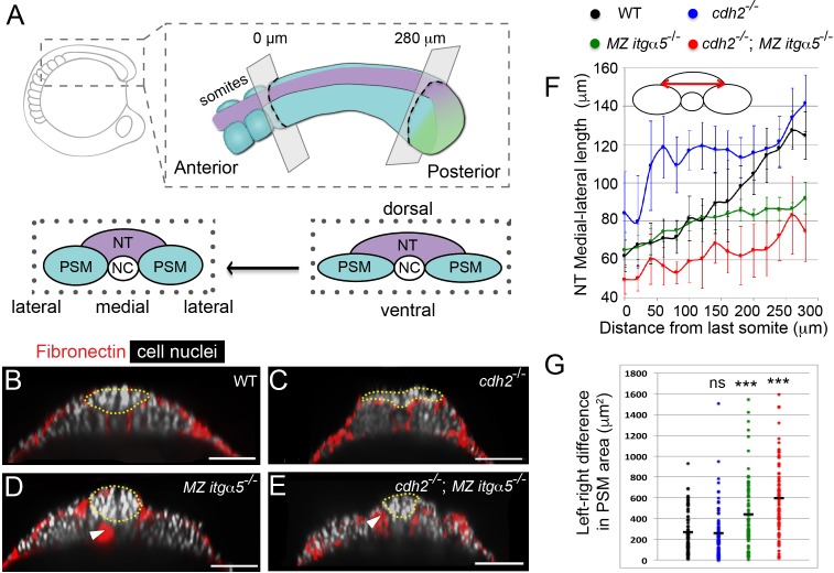
Figure 1.

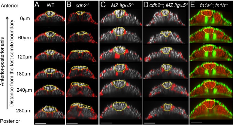
Figure 1—figure supplement 1. Reduction of cell-ECM interactions leads to precocious neural tube convergence and rescues cdh2 mutant neural tube convergence defects.

Figure 1—figure supplement 1.

Each column shows a series of transverse sections in 12–14 somite stage embryos along the anterior-posterior axis of the tailbud starting from the last somite boundary (0 μm) in WT (A), cdh2-/- mutants (B), MZ itgα5-/- mutants (C), cdh2-/-; MZ itgα5-/- mutants (D), and fn1a-/; fn1b-/- mutants. (A–D) Immunostaining for FN (red) and nuclei labeling (grey). (E) F-actin labeling (green), and nuclei labeling (red). Yellow dotted lines delineate neural tube contours. Dorsal is to the top. Scale bars = 70 μm.

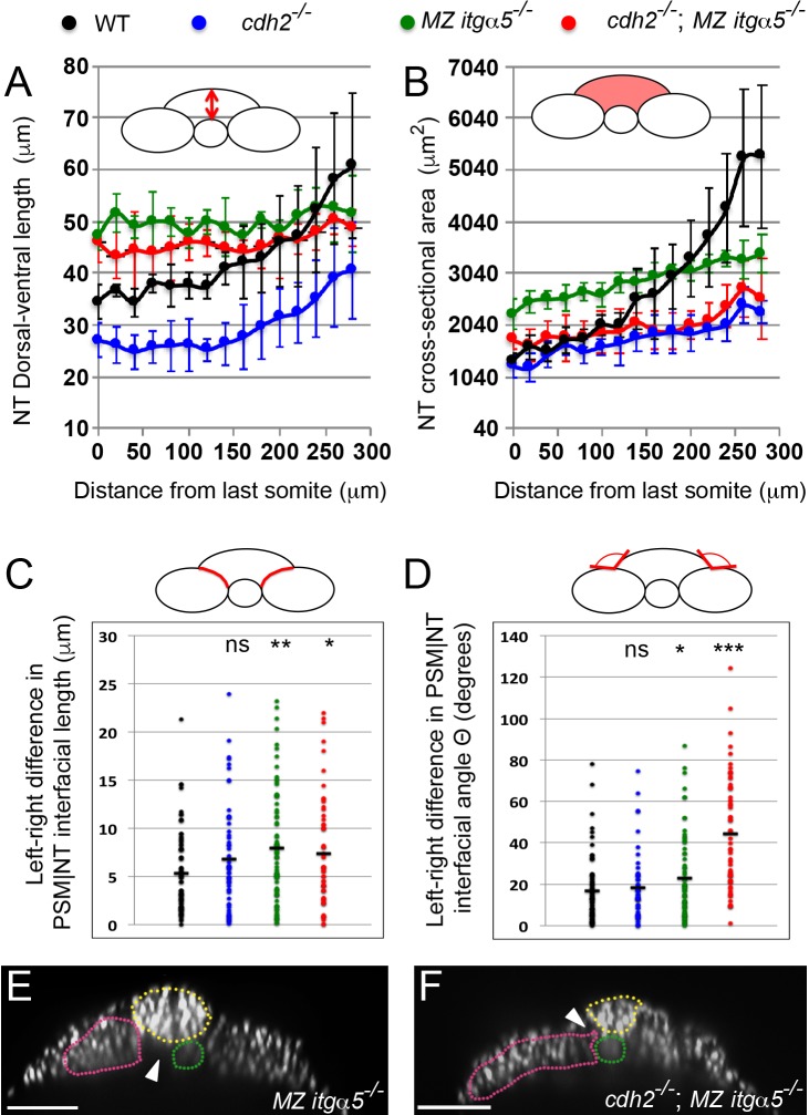
Figure 1—figure supplement 2. Reduction of cell matrix interactions provokes a precocious neural tube convergence and generates left-right asymmetries in the PSM|NT interfacial length and angle.

Figure 1—figure supplement 2.

(A, B) Quantification of the dorsal-ventral length (A, as indicated by red double arrow) and the cross-sectional area (B, as indicated by the red area) of the neural tube along the anterior-posterior axis starting from the last somite boundary (0 μm). Measurements were performed on transverse sections taken every 20 μm. Dots represent means and error bars represent SD. Sample size n = 10 PSMs on five embryos for each genotype. (C, D) Quantification of left-right asymmetry in PSM|NT interfacial length (C) and PSM|NT interfacial angle (D). Each dot denotes an absolute difference in left and right PSM|NT interfacial length or angle in a transverse section. Sample size n = 75 sections from five embryos for each genotype. ***p<0.0005, **p<0.005, *p<0.05, via T-test. (C) cdh2-/- vs WT, p=0.095; MZ itgα5-/- vs WT, p=1.8e-3; MZ itgα5-/-; cdh2-/- vs WT, p=0.23. (D) cdh2-/- vs WT, p=0.57; MZ itgα5-/- vs WT, p=0.046; MZ itgα5-/-; cdh2-/- vs WT, p=1.56e-9. (E–F) Transverse sections for MZ itgα5-/- (E), and cdh2-/-; MZ itgα5-/- (F) identical to those presented in Figure 1D and Figure 1E but showing only the nuclei signal to highlight tissue detachments (arrowheads) between the notochord (green), the PSM (pink) and the neural tube(yellow).