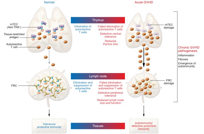
Figure 1. Model of impaired tolerance in GVHD pathogenesis.
Acute GVHD simultaneously damages nonhematopoietic cells enforcing central and peripheral tolerance in the thymus and lymph nodes. In the thymus, mTECs ectopically express a broad range of tissue-restricted antigens under the control of the autoimmune regulator AIRE. mTECs promote the negative selection of autoreactive T cells and development of central tolerance. In the periphery, lymph node FRCs also show promiscuous expression of tissue-restricted antigens and facilitate the suppression of mature autoreactive T cells, thereby maintaining peripheral tolerance. Acute GVHD targets both mTECs in the thymus and FRCs in lymph nodes, thereby creating a dangerous situation in which large numbers of autoreactive T cells are left to sustain tissue damage during chronic GVHD. TRA, tissue-restricted antigen; GVHD, graft-versus-host disease; mTECs, medullary thymic epithelial cells; AIRE, autoimmune regulator; FRCs, fibroblastic reticular cells.

