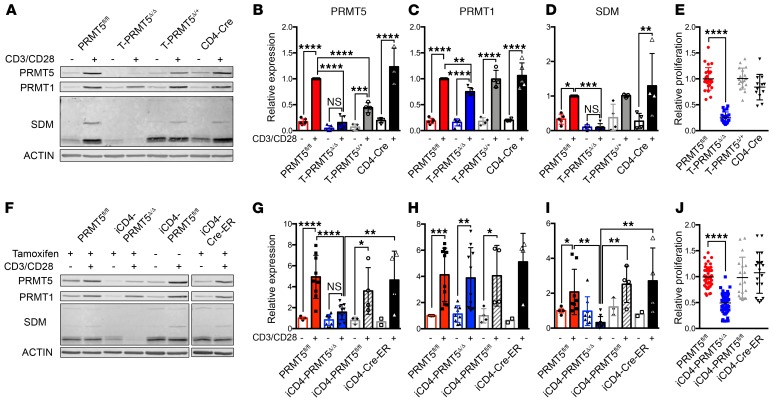
Figure 3. Prmt5 deficiency suppresses T cell proliferation.
Whole spleen CD4+ T cells from (A–E) T-PRMT5Δ/Δ or (F–J) iCD4-PRMT5Δ/Δ mice were isolated and activated with anti-CD3/CD28. Cells were collected for immunoblotting directly ex vivo and after 48 hours of anti-CD3/CD28 activation and (A and F) analyzed by immunoblot. Lysates were run on the same gel but are noncontiguous in F. Bands detected by (B and G) anti-PRMT5, (C and H) anti-PRMT1, and (D and I) PRMT5’s symmetric dimethylation mark (anti-SYM10 antibody) were quantified using ImageStudio software. Data are pooled from at least 3 representative independent experiments (n = 5–7 independent mice). (E and J) Proliferation of CD4+ T cells was analyzed by 3H-thymidine incorporation and expressed as a relative proliferation ratio to the resting PRMT5fl/fl control condition. Data include at least 3 independent experiments (n = 5–12 mice/group). Two-way ANOVA followed by Sidak’s multiple-comparisons test (B–D and G–I) or 1-way ANOVA followed by Dunnett’s multiple-comparisons test (E and J). *P < 0.05, **P < 0.01, ***P < 0.001, ****P < 0.0001. Graphs display mean ± SD.

