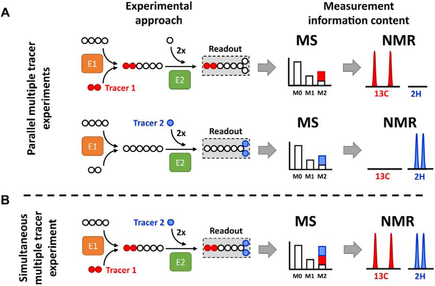
Figure 1. Application of multiple stable isotope tracers.

Example metabolic network consisting of two enzymatic reactions (E1 and E2). Tracer 1 provides two 13C atoms, while tracer 2 provides two 2H atoms. (A) Parallel experimental setup: tracer 1 and 2 are administered in two separate biological experiments generating two separate datasets. Therefore, individual MIDs and NMR spectra contain labeling information originating from each single tracer. (B) Simultaneous experimental setup: both tracers are administered in the single biological experiment and generate a single dataset. Labeling from tracer 1 and 2 becomes convoluted in the MID and is difficult to interpret without mathematical modeling. In contrast, NMR detection is nucleus-sensitive and can distinguish between signals originating from 2H and 13C atoms.
