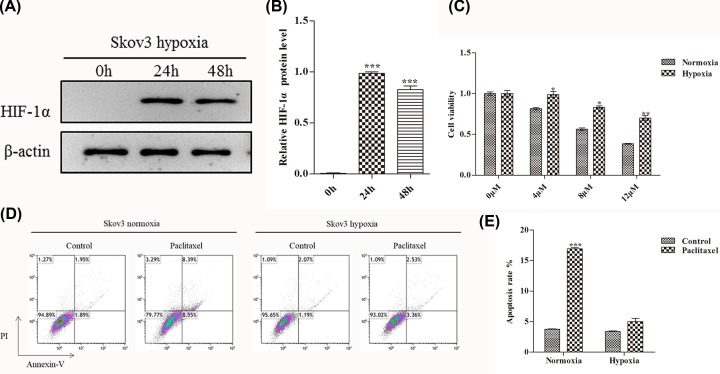
Figure 1. Hypoxia increases cell viability and reduces apoptosis in OC.
(A) WB analysis the expression of HIF-1α protein in Skov3 cells when hypoxia treatment for 0, 24 and 48 h. (B) Quantify the protein bands of the HIF-1α protein (O.D. ratio over β-actin). (C) Cell viability of Skov3 cells was measured by MTT assay. Skov3 cells were treated with 0, 4, 8 and 12 μM paclitaxel under the condition of hypoxia or normoxia. (D and E) Flow cytometric analysis of skov3 cell apoptosis rate when treated with paclitaxel (4 μM, 48 h) under the condition of hypoxia or normoxia. Data are shown as mean ± SEM (n = 3). Asterisks indicate significant differences from the control (*, P <0.05; **, P <0.01; ***, P <0.001, Student’s t-test).

