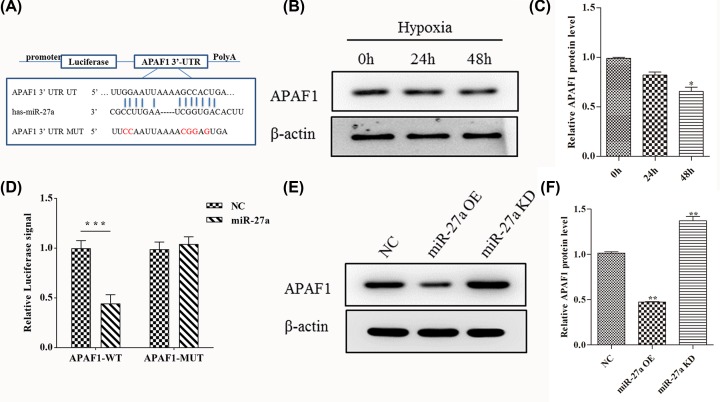
Figure 3. MiR-27a regulates gene expression of its target APAF1.
(A) 3′-UTR base pairing diagram of miR-27a and APAF1. Replacement of Guanine base with Cytosine (G to C) or replacement of Cytosine bases with Guanine (C to G) can also be used for the construction of mutant reporter. (B) The expression of APAF1 protein in Skov3 cells by Western blot when hypoxia treatment for 0, 24 and 48 h. (C) Quantify the protein bands of the HIF-1α protein (O.D. ratio over β-actin). (D) Cells were co-transfected with miR-27a mimics and a luciferase reporter containing a fragment of the APAF1 3′-UTR harboring either the miR-27a binding site (APAF1-3′-UTR-WT) or a mutant (APAF1-3′-UTR-MUT). (E and F) The APAF1 protein expression in the miR-27a OE and miR-27a KD cells by Western blot analysis. Data are shown as mean ± SEM (n = 3). Asterisks indicate significant differences from the control (*P <0.05; **P <0.01; ***P <0.001, Student’s t-test).

