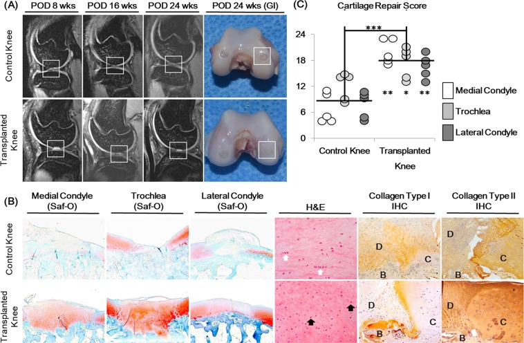
Figure 5.
Cartilage repair analysis of 2-week cultured cartilage gels transplanted in a non-human primate cartilage defect model. (A) MRI images of femoral condyles taken at 8, 16 and 24 weeks after surgery along with final gross images taken at 24 weeks showed continued filling of the cartilage defect. (B) Histological analysis of cartilage defects 24 weeks after surgery showed better cartilage repair in cartilage gel groups, compared to defect only control groups. Repaired cartilage of the cartilage gel group showed lacunae formation (black arrow), and absence of cell clustering present in the control group (white arrow). Repaired cartilage expressed similar levels of collagen type I and II to surrounding hyaline cartilage. (C) Dot plot of cartilage repair score results. Cartilage repair scores were significantly better overall and in each anatomic location of the defect compared to the control group. Statistical analysis done with Mann-Whitney test (*p < 0.05, **p < 0.01, ***p < 0.001). Magnification x15, x100, x200. GI; Gross image, H&E; Hematoxylin and eosin, SO; Safranin-O, IHC; Immunohistochemistry, D; Defect, C; Native Cartilage, B; Bone.

