Figure 6.
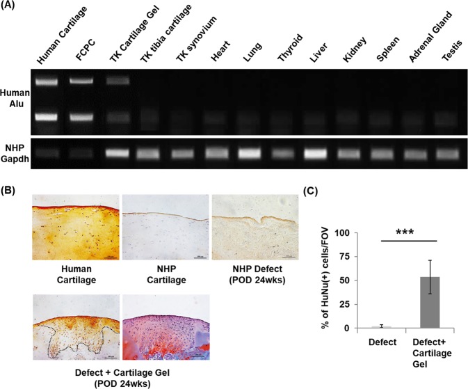
Cell distribution of transplanted cartilage gels in a non-human primate cartilage defect model after 24 weeks. (A) Representative RT-PCR result bio-distribution using human specific Alu sequence. Human Alu was detected in the transplanted cartilage tissue and was absent in adjacent tibial cartilage, synovium, and other vital organs. (B) Cell tracking using human anti-nuclear antibody. Immunohistochemical staining results showed that cartilage gels of human origin remained within the defect after 24 weeks. Safranin-O slide shown as reference for repair site. (C) Semi-quantification of immunohistochemistry results showed over 50% of human anti-nuclear antibody (+) cells within the defect. Statistical analysis done with Mann-Whitney test (*p < 0.05, **p < 0.01, ***p < 0.001). Magnification x40. FCPC; fetal cartilage progenitor cells, TK; transplanted knee, NHP; non-human primate, HuNu; human anti-nuclear antibody, FOV; field of view (x40).

