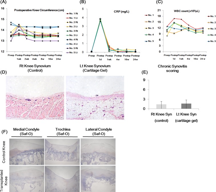
Figure 7.
Postoperative inflammation analysis of non-human primate cartilage defect model after cartilage transplantation: Right (Rt) knees received defect surgeries as controls, while left (Lt) knees received cartilage gel transplantation. (A) Knee circumference was measured before and after transplantation in all five animals to assess indirect inflammation on the joints. Note that knee circumferences returned to baseline in all groups by postop week 4. (B) WBC count was measured from whole blood before and after transplantation to assess inflammation. Note that no leukocytosis/ leukopenia was observed in all groups beyond postop week 4. (C) C-reactive protein level was measured from whole blood before and after transplantation to assess inflammation. Note that all animals achieved levels below 1 mg/L by postop week 1. (D) Histology (H&E) of synovium. None to mild inflammatory reactions were observed in all synovium, with no difference between controls and cartilage gel transplantation groups (E). Overall, signs of rejection, or chronic inflammation were not present in all animals. (F) IHC stain for inflammatory marker CD45 was performed. Statistical analysis done with Mann-Whitney test (*p < 0.05, **p < 0.01, ***p < 0.001). Magnification x100, x40.

