Figure 1.
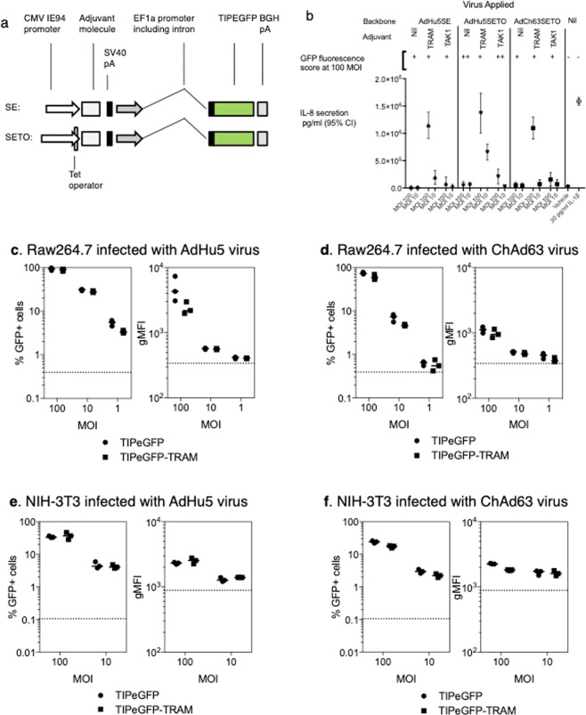
TRAM-induced inflammatory signaling and adenovirus expression cassettes. (a) AdHu5 and ChAd63 adenoviruses were constructed by incorporating expression cassettes (illustrated) into the deleted E1 region. Two cassettes were studied, SE and SETO, the latter featuring repression of CMV promoter function during viral growth. All viruses expressed a model antigen (TIPEGFP) driven by the human EF1α promoter. (b) TRAM expression induces IL-8 response in vitro HeLa cells were either exposed to adenoviruses at an MOI of 10 or 100, or stimulated with IL1β, or exposed to vehicle (Optimem medium) for one hour. 36 hours after infection, antigen expression was assessed by microscopy and IL-8 secretion measured by EIA. (c–f) Co-expression of mouse TRAM does not increase the antigen transgene expression in mouse cell lines. Raw246.7 (c,d) or NIH-3t3 (e,f) cells were infected with the AdHu5 (a,e) or ChAd63 (d,f) at an MOI of 100, 10 or 1. Cells were harvested 24 hours later and the level of GFP expression measured by flow cytometry. Graphs represent the percentage of infected cells (GFP+) or the geometric mean fluorescence intensity (gMFI) of the GFP signal, each dot represents a replicate well with lines representing the median response.

