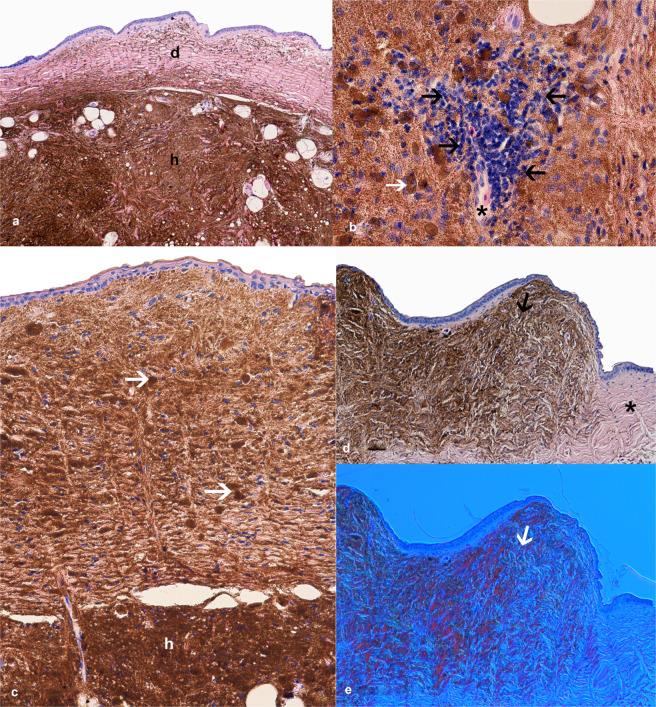
Figure 3.
Histopathology of the leopard gecko skin iridophoroma. (a–d) H&E. (e) Nomarski contrast (DIC). (a) Large mass of iridophores in hypodermis (h) and scattered clusters of iridophores in dermis (d), regular collagen fibre arrangements were noted. (b) Infiltration of lymphohistiocytic cells (black arrows) around a skin capillary (black asterisk), atypical oval shaped iridophore with eccentrically located nucleus and brown cytoplasm (white arrow). (c) Tumour cells present both in the epidermis, dermis and hypodermis (h), round and spindle shaped cells filled with guanine crystals (white arrows), regular collagen fibre arrangement. (d) Overview of the affected skin with a tumour lesion (black arrow) and healthy margin without iridophores and other chromatophores (black asterisk). (e) Nomarski contrast used to confirm the presence of iridophores in the skin sections; note the characteristic change in colour of light-reflecting cells (white arrow).

