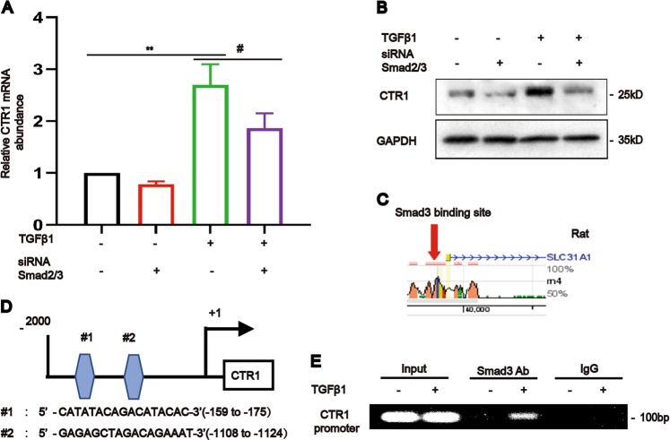
Fig. 2. TGF-β1 upregulated CTR1 via Smad3 binding to the CTR1 promoter.
a, b Real-time PCR and western blotting and analyses showed the expression of CTR1 in NRK-49F cells after the simultaneous knockdown of Smad2 and Smad3 and treatment as indicated (n = 5). c, d Binding protein prediction in the rat CTR1 promoters using two evolutionarily conserved predicted binding sites (http://ecrbrowser.dcode.org/ and http://jaspardev.genereg.net/) for Smad3. Note that the NRK-49F cell line used in this study originated in rats. e Chromatin immunoprecipitation (ChIP) assay followed by PCR showed that Smad3 physically binds to the CTR1 promoter in response to TGF-β1 after 24 h (n = 5). Data represent the mean ± SEM. **P < 0.01 versus blank control cells. #P < 0.05 versus TGF-β1-treated cells.

