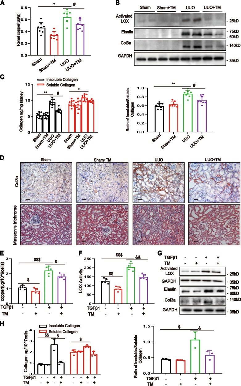
Fig. 5. Chelating copper decreased LOX activity and ameliorated renal fibrosis.
UUO or sham-operated rats were randomly divided into the following four groups: (1) sham rats treated with PBS; (2) sham rats treated with TM (1.2 mg/kg); (3) UUO rats treated with PBS; and (4) UUO rats treated with TM. a Copper concentrations of kidneys were determined by ICP-MS (n = 8). b Western blotting showing actived LOX, elastin and col3a expression in rat kidneys (n = 6). c The kidney insoluble collagen, soluble collagen and their ratio were determined (n = 8). d Representative images of col3a immunostaining and Masson’s staining of kidney sections (n = 5). Original magnification, ×200. Bar = 50 μm. e ICP-MS analysis of copper concentrations in NRK-49F cells at 24 h with or without TM and TGF-β1 treatment (n = 5). f LOX activity in cell culture medium assay was determined using the Fluorometric Lysyl Oxidase Assay Kit (n = 5). g Western blotting showing the expression of activated LOX, elastin and col3a (n = 3). h Microwave-assisted acid digestion showing the insoluble and soluble collagen in NRK-49F cells with or without treatment of TM and the ratio of insoluble to soluble collagen were calculated (n = 3). Data represent the mean ± SEM. *P < 0.05, **P < 0.01 versus sham-treated rats. #P < 0.05 versus solvent control-treated rats with UUO. $P < 0.05, $$P < 0.01, $$$P < 0.001 versus nontreated cells. &P < 0.05, &&P < 0.01 versus TGF-β1-treated cells.

