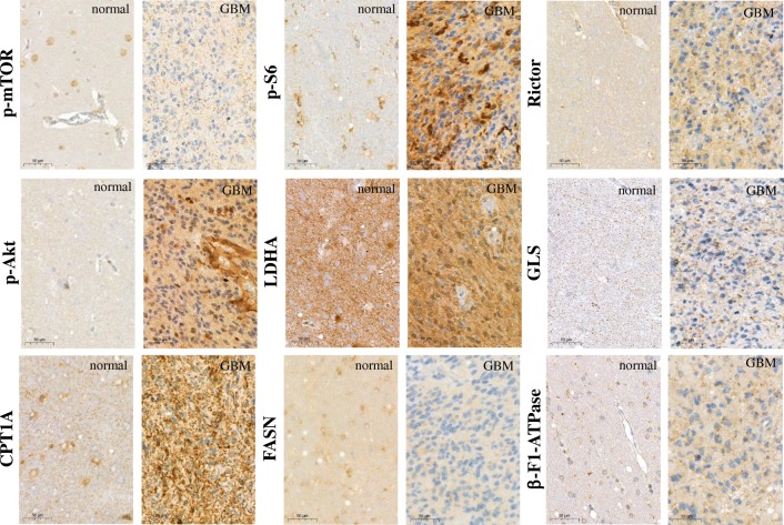
Fig. 2.
Representative immunostainings with different metabolic markers in human high-grade gliomas and normal brain tissues. The antibody-stainings were developed with DAB substrate (brown) and counterstained using haematoxylin. mTOR activity was characterised by p-mTOR, p-S6, Rictor, p-Akt stainings, other metabolic activity correlates to LDHA, GLS, CPT1A/FASN and β-F1-ATPase

