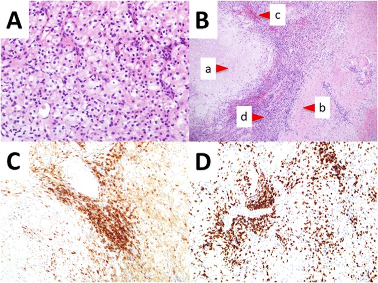
Fig. 2.
A HE staining of renal cell carcinoma, B biopsy imaging showing no residual tumor of the primary renal mass, and C immunostaining of CD4 and D CD8. The primary renal mass was completely replaced by a necrosis, b fibrosis, hyalinization, c hemorrhage, and d macrophages. (a × 400, b × 100, c × 200, d × 200)

