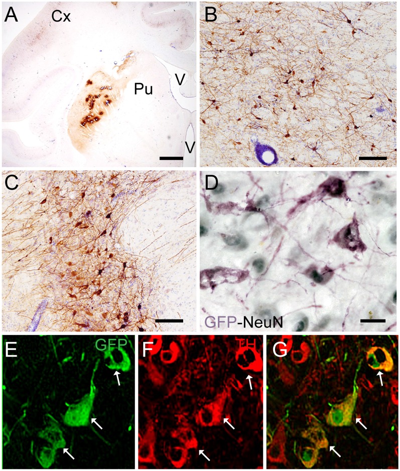
Figure 1.
Coronal sections of animals injected with canine adenovirus type (CAV)-GFP in the left putamen. (A) Low magnification of immunohistochemistry (IHC) against GFP and Nissl counterstained of the injected putamen; (B) higher magnification of IHC against GFP and Nissl counterstained of the injected thalamus; (C) IHC against GFP and Nissl counterstained of the substantia nigra (SN) of the injected hemisphere; (D) IHC against GFP (pink) and NeuN (black) in the SN of the injected hemisphere; and in the contralateral SN (E) immunofluorescence (IF) against GFP (green; F) IF against TH (red; G) merge of (E,F). White arrows denoted TH+/GFP+ cells. Scale bars: (A) 1 mm; (B,C) 100 μm; (D) 10 μm; (E) 5 μm.

