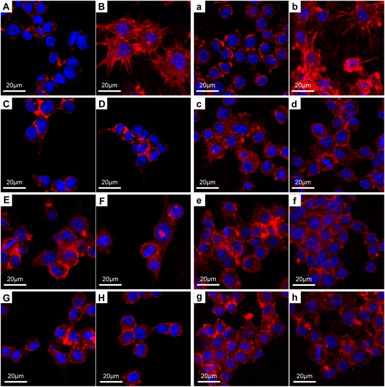
Figure 9.
CLSM images of RAW 264.7 cells after culturing for 1 and 3 days on different samples.
Notes: Uppercase letters indicate the day 1 images, lowercase letters indicate the day 3 images: (A, a), Ti; (B, b), Ti surface with 1 μg/mL LPS treatment; (C, c), 5VZn surface with 1 μg/mL LPS treatment; (D, d), 5V surface with 1 μg/mL LPS treatment; (E, e), 15VZn surface with 1 μg/mL LPS treatment; (F, f), 15V surface with 1 μg/mL LPS treatment; (G, g), 25VZn surface with 1 μg/mL LPS treatment; (H, h), 25V surface with 1 μg/mL LPS treatment.
Abbreviations: CLSM, confocal laser scanning microscope; LPS, lipopolysaccharides.

