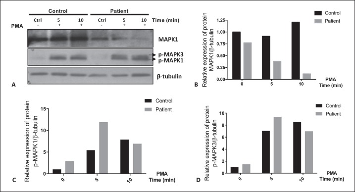
Fig. 3.
A-D MAPK1 protein from activated PBMCs is decreased in the patient with 22q11.2DDS. A PBMCs from the patient with 22q11.2DDS and the control volunteer were unstimulated or stimulated with PMA for 5 and 10 min. MAPK1/MAPK3 protein was detected using the total ERK2/ERK1 mouse monoclonal antibody and phospho-specific rabbit polyclonal antibodies. A secondary antibody labeled with horseradish peroxidase was used for detection by chemoluminescence. Densitometry of total MAPK1 (B), phosorylated MAPK1 (C), and MAPK3 (D) were normalized to β-tubulin.

