Figure 5.

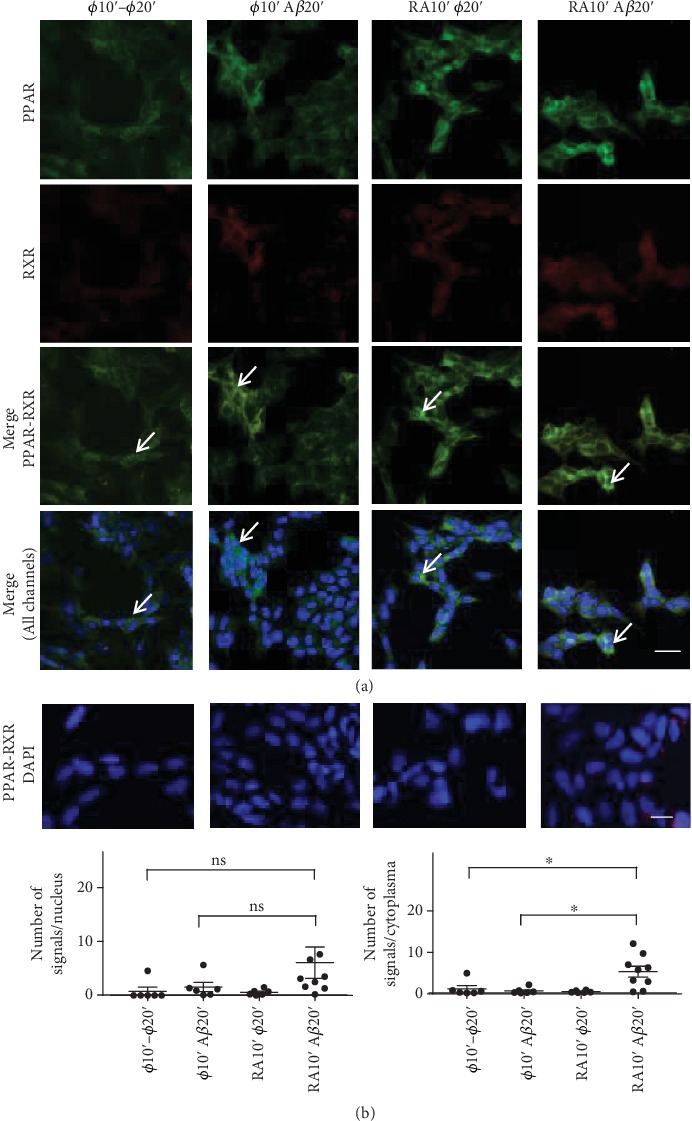
Neuroprotection experiments carried out with SH-SY5Y cells to show the activation of RA-dependent pathways by a 10 min RA treatment or not (ø), followed by a 20 min treatment with Aβ or not (ø10′–ø20′, ø10′–Aβ20′, RA10′–ø20′, and RA10′–Aβ20′). (a) By immunofluorescent cytochemistry, it was shown that PPARβ/δ (green) increased in the cytoplasm with the RA–Aβ treatment, whereas a cytoplasmic increase of the RXRα/β/γ (red) could not be observed. However, sites of colocalisation (white arrows, green-yellow signals) could be detected (Merge, PPAR-RXR, or all channels). Signals were too faint in the cell nucleus (DAPI). (b) Duolink™ proximity ligation assay was used to determine PPARβ/δ and RXRα/β/γ heterodimerization under the same conditions as in (a). A significant increase of PPAR-RXR signals was observed with the RA–Aβ treatment only in the cytoplasm when compared to the ø–ø (p = 0.0125) or ø–Aβ (p = 0.0145) treatments. Only a nonsignificant (ns) increase was observed in the cell nuclei. The number of pictures analysed is 6 to 9. Similar experiments for RARα and RXRα/β/γ resulted in no significant differences. Scale bar for (a): 25 μM and for (b): 13 μM.
