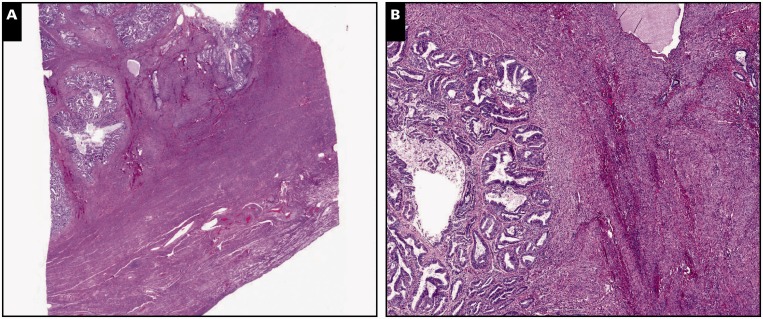
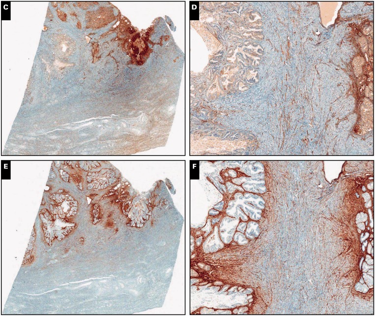
Image 1.
Myoinvasive endometrial adenocarcinoma, endometrioid type, in a background of adenomyosis, scanning (A, C, and E; ×5) and ×30 (B, D, and F) views. A and B, Myometrial invasion is present on the left side of the tissue section, characterized by packed neoplastic glands dissecting myometrium in a pushing/expansile pattern (H&E).
C and D, By immunohistochemistry, interferon-induced transmembrane protein 1 is negative around myoinvasive carcinoma, showing expression only in stroma of the endometrium and adenomyosis. E and F, CD10 immunohistochemistry shows strong and diffuse staining around both invasive and noninvasive carcinoma.


