Image 4.
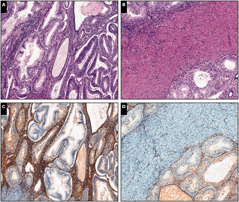
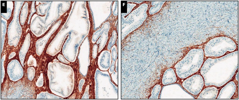
Endometrial adenocarcinoma, endometrioid type, extending to adenomyosis, ×80 (A, C, and E) and ×100 (B, D, and F) views. A and B, Residual normal endometrial glands and stroma are identified in the periphery of and in between neoplastic glands (H&E). C and D, Diffuse cytoplasmic interferon-induced transmembrane protein 1 expression in the endometrial stroma.
E and F, A similar pattern is observed with CD10.


